Very frequently we encounter clinical cases dental and dentomaxilar disharmonies that cannot be remedied, or treated, only by surgery or orthodontic or prosthetic treatments.
Only the coordinated combination of the interdisciplinary treatments can lead to an entirely aesthetic and stable result over time. In the case we present, that of a patients treated in the Sirona clinic, who came to us with the desire to improve the unsightly appearance of the “midline space” (diastema), we used a multidisciplinary approach.
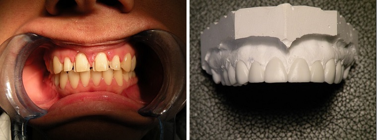
Namely, on the orthodontic plan, it was required to reduce the diastema but not to remove it completely, since the front teeth showed a congenital lack of dental tissue, the teeth being much smaller in the tri-dimensional norm than they should have, proportionally comparing themt o the lateral teeth and the lower central teeth (from the lower jaw). This can be calculated very precisely thrugh an analysis and measurement of the gypsum model, applying the “BOLTON” formula.

In case of not meeting this analysis and in the lack of dental tissue, most times we proceed to the total closure of the space through the orthodontic methid called mesialization of the teeth and narrowing of the transverse dimension of the jaw. Most often the result is unsatisfactory for the patient because it produces the appearance of many small and crowded teeth but also a flattening of the face profile and lips. Exemplified on the simulation model in fig.5.
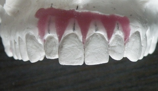
The Bolton formula indicates us exactly, in tenths of a millimeter, what width each tooth should have. So we orthodontically aligned the teeth without completely closing the spaces between them, but keeping the exact size that a correctly formed tooth shpuld have.
The surgical part consisted of the excision of the upper lip frenulum which was inserting (it originated) very deep exactly between the two central incisors. This was we stopped the traction in that area of the lip muscles and it allows the formation of bone and the stabilization of the teeth position obtained with the orthodontic appliance.
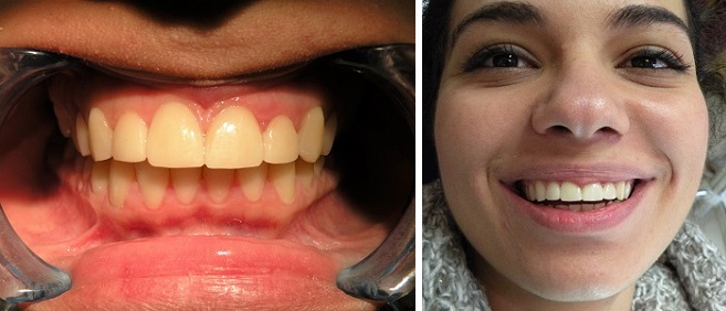
The dental tissue deficit was completed in the last phase by preparing the front teeth 12,11,21,22 and prosthesis thereof with ceramic veneers. The presentation of a “wax-up” early into the treatment and the veneers in the final position: Fig. 6, 7.
Thus, combining the three treatments we were able to obtain through a professional approach an outcome that fully satisfied the patient both aesthetically and functionally. Fig. 8, 9.